The fascinating architecture of the lumbar spine

The lumbar spine (LWS) carries our body weight daily and simultaneously allows for amazing freedom of movement. It manages these seemingly contradictory tasks thanks to its special structure. But why are the five lumbar vertebrae significantly more massive than the vertebrae of the thoracic or cervical spine?
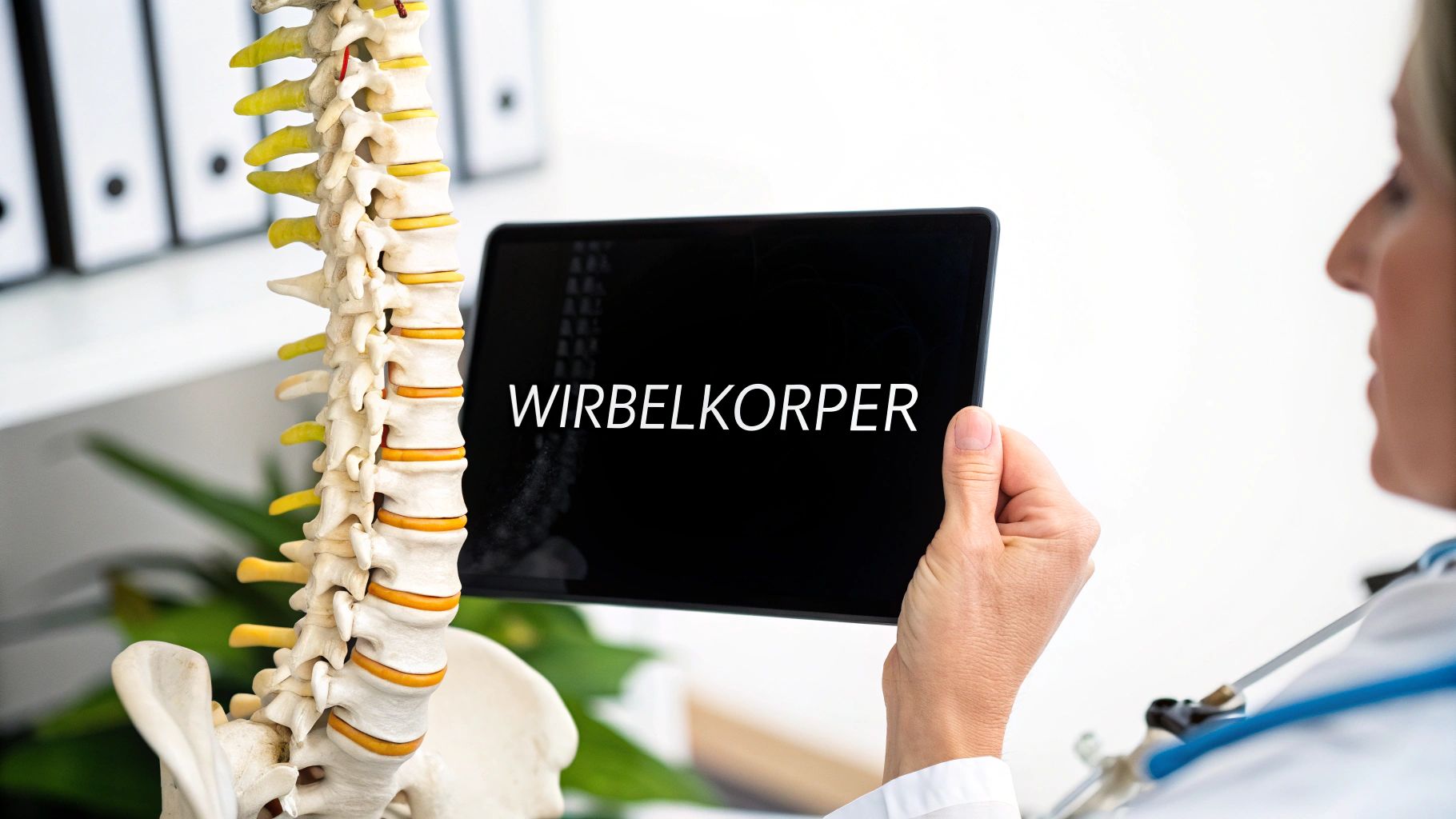
The answer lies in our upright posture. As the largest and strongest vertebrae of the spinal column, the lumbar vertebrae bear the main load of the upper body. This adaptation allows us to walk and stand upright in the first place. The lumbar spine is therefore essential for the stability of the body, but is heavily stressed by the upright posture. This often leads to back problems. In Germany, almost half of all early retirements are due to spinal problems. The annual costs for treatments and lost work time amount to over 20 billion euros. The lumbar spine consists of five vertebrae, between which intervertebral discs are located. It is physiologically curved forward and forms a so-called lordosis angle. More information on the strain on the lumbar spine can be found here. The high load makes the lumbar spine susceptible to wear and tear and injuries. A healthy lumbar spine in everyday life starts with a balanced lifestyle that also promotes inner peace .
The Building Blocks of the Lumbar Spine
Each lumbar vertebra is composed of various components:
- Vertebral body: The cylindrical vertebral body is the load-bearing part of the vertebra and is particularly resistant to compressive stress.
- Vertebral arch: It encloses the spinal cord and forms the vertebral canal.
- Facet Joints: These small joints connect the individual vertebrae and allow movement in different directions.
- Vertebral Processes: They serve as attachment points for muscles and ligaments.
The Lordosis: A Biomechanical Marvel
The natural curvature of the lumbar spine, the so-called lordosis, is another important feature. This forward curvature acts like a shock absorber. It distributes the load evenly across the spine and thus protects the vertebral bodies.
In addition to the lordosis, the S-shape of the entire spine contributes to stability and flexibility. This double S-shape is a masterpiece of biomechanics. It allows us to walk upright, run, jump, and turn. Even small changes in the anatomy of the lumbar spine can have far-reaching consequences and lead to pain and limited movement. Therefore, it is important to understand the anatomy of the lumbar spine and to support it through targeted exercises and a healthy lifestyle.
Intervertebral Discs & Ligaments: The Silent Heroes of the Back
Our lumbar spine is a complex marvel. We often only think about its most important components when pain occurs. Intervertebral discs and ligaments - the silent heroes of our back - ensure stability and flexibility. But how do they actually work?
The Intervertebral Discs: Shock Absorbers for the Spine
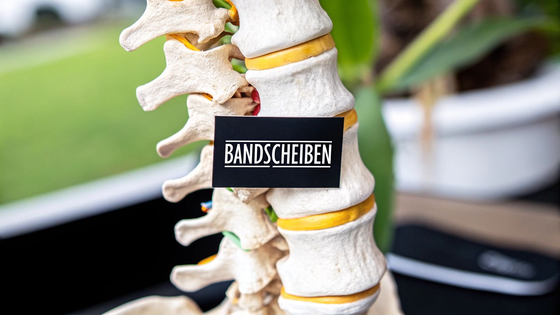
The intervertebral discs are the flexible connections between the vertebral bodies. They consist of two main parts: the gelatinous core (Nucleus pulposus), a gel-like center, and the fibrous ring (Anulus fibrosus), which surrounds the core. Together they act like a perfect shock absorber. The gelatinous core absorbs the load and distributes the pressure evenly to the fibrous ring. This prevents the gelatinous core from slipping.
The following table illustrates the structure, function, and common changes of the intervertebral discs in the lumbar spine.
Lumbar Intervertebral Discs: Comparison of the Structural Components and Functions of the Intervertebral Discs
| Component | Structure | Function | Common Changes |
|---|---|---|---|
| Nucleus Pulposus | Gel-like substance, rich in water | Absorbs shocks and distributes pressure | Fluid loss, dehydration |
| Annulus Fibrosus | Multiple layers of fibrous cartilage | Encloses the nucleus pulposus, stabilizes the intervertebral disc | Tears, protrusions, herniated disc |
The table clearly shows how important the water content of the nucleus pulposus is for the function of the intervertebral disc and which problems can arise from its loss.
The Ligaments: The Natural Corset
Besides the intervertebral discs, the ligaments are essential for the stability of our lumbar spine. They form a complex network that connects and supports the vertebrae – like a natural corset. At the same time, the ligaments allow the mobility we need in everyday life.
These ligaments function similarly to elastic bands, holding the vertebrae securely together while allowing movement. They provide support to the spine and prevent excessive movements.
Age-Related Changes and Prevention
With increasing age, intervertebral discs and ligaments change. The nucleus pulposus loses fluid, the annulus fibrosus can tear, and the ligaments lose elasticity. These changes are normal and a reason why back problems increase with age. You might be interested in: How to master...
To maintain the functionality of intervertebral discs and ligaments for as long as possible, prevention is important. Regular exercise, targeted training of the back and abdominal muscles, and an ergonomic posture in everyday life relieve the lumbar spine and protect its structures. This helps prevent back pain and maintain your mobility and quality of life.
The powerful muscle-nerve orchestra of the lumbar spine
Our lumbar spine is a complex structure. It is surrounded and controlled by a finely tuned system of muscles and nerves. You can imagine it like an invisible support corset that allows us a wide range of movements while ensuring stability in the lower back. Let's take a look at the most important components of this fascinating orchestra.
The Muscles: Supporting Pillars of the Back
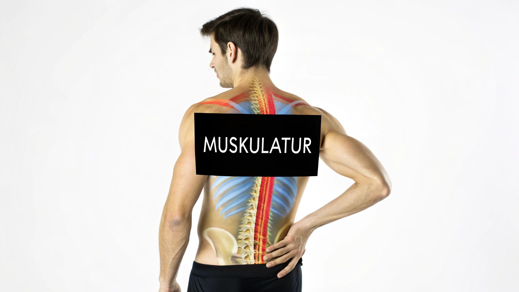
Two muscle groups are particularly important for the anatomy of the lumbar spine: the back extensor (M. erector spinae) and the abdominal muscles. The back extensor runs along the spine and is primarily responsible for straightening and backward bending of the upper body. Thanks to it, we can stand upright and lift objects.
The abdominal muscles act as antagonists and ensure balance. By bending the lumbar spine forward and supporting the abdomen, they stabilize the torso. A strong abdominal muscle corset therefore significantly relieves the lumbar spine.
The Nerves: The Body's Information Network
The nerves of the lumbar spine control the muscles and transmit sensory information. They originate from the spinal cord as nerve roots L1 to L5 and unite to form the lumbar plexus. This is a complex nerve network that supplies the legs and lower back.
Imagine these nerve pathways as the signal cables of our body. Information constantly flows from the brain to the muscles and from the skin and joints back to the brain. Even the smallest nerve compressions, for example, due to a herniated disc, can disrupt these signal pathways and lead to severe pain.
Interaction for a healthy back
The harmonious interaction of muscles and nerves is essential for a healthy and functional lumbar spine. An imbalance can lead to pain, limited movement, and postural problems.
Targeted training for back and abdominal muscles is therefore essential. Strengthening and stretching exercises, tailored to the anatomy of the lumbar spine, stabilize the muscles and relieve the nerves. Even in everyday life, we can protect our lumbar spine and prevent pain through back-friendly posture and by avoiding improper strain. Those who understand this complex interplay can actively promote the health of their lumbar spine and improve their quality of life.
Understanding Movement: The Biomechanics of the Lumbar Spine
Our lumbar spine is a complex structure that enables us to perform a variety of movements every day. It is much more than just a static element of our body. From simple bending to twisting – the biomechanics of the lumbar spine is a fascinating interplay of various components: bones, joints, muscles, and nerves work together perfectly here. But why is our lumbar spine better suited for some movements than others?
Flexion and Extension: Bending and Stretching
The lumbar spine is ideally designed for flexion (forward bending) and extension (backward bending) . The shape of the vertebral bodies and the special arrangement of the facet joints make these movements possible. For example, when we bend forward to tie our shoes, the vertebral bodies glide smoothly past each other. At the same time, the intervertebral discs are compressed at the front and stretched at the back.
The opposite is true for backward bending, such as stretching after getting up. Here, the vertebral bodies approach each other in the posterior area, which stretches the intervertebral discs at the front and compresses them at the back. Flexion and extension are thus part of the natural range of motion of our lumbar spine.
Rotation: Rotations and their Limits
Compared to flexion and extension, the rotation (rotational movement) of the lumbar spine is significantly more restricted. The position of the facet joints limits the rotational movement to ensure the stability of the entire spine. Try rotating your upper body while keeping your pelvis fixed. You will notice that the movement in the lumbar spine is limited. The majority of rotation occurs in the thoracic spine.
Pressure Conditions: Influence of Posture and Activity
The load on the lumbar spine varies greatly – depending on our posture and the activities performed. When lying down, the pressure on the intervertebral discs is lowest. When standing, this pressure increases noticeably. The load is particularly high when lifting heavy objects, especially in a bent posture. This puts enormous pressure on the intervertebral discs, which can lead to damage to the discs and vertebrae .
The following table shows the normal ranges of motion of the lumbar spine in different age groups. It illustrates how mobility changes over the course of a lifetime.
Lumbar Spine Range of Motion Statistical data on normal ranges of motion in different age groups
| Direction of Movement | Standard Value (20-30 years) | Standard Value (40-50 years) | Standard Value (60+ years) |
|---|---|---|---|
| Flexion (Forward Bending) | 40-60° | 30-50° | 20-40° |
| Extension (Backward Bending) | 20-35° | 15-25° | 10-20° |
| Rotation (Twisting) | 15-20° | 10-15° | 5-10° |
As the table shows, the mobility of the lumbar spine decreases with increasing age. This underscores the importance of regular exercise and back-friendly behavior.
Back-Friendly Movement in Everyday Life
Understanding the biomechanics of the lumbar spine helps us to develop and apply back-friendly movement patterns in everyday life. Maintain an upright posture and avoid twisting movements under load. Lift heavy objects by bending your knees and keeping your back straight to distribute the load evenly. Regular training of the back and abdominal muscles strengthens the natural support corset. With these simple tips, you can actively contribute to the health of your lumbar spine and prevent back pain.
When Anatomy Causes Problems: Pathological Changes
The lumbar spine is a complex structure that supports us and enables movement every day. However, its complicated structure makes it susceptible to various diseases. Genetic predisposition, age, and improper strain – the factors that can lead to pathological changes are manifold. And the consequences can be far-reaching: pain, limited mobility, and a significantly reduced quality of life.
Herniated Disc: When the Nucleus Pulposus Protrudes
A herniated disc is one of the most common diseases of the lumbar spine. In this condition, the annulus fibrosus of the intervertebral disc ruptures and the nucleus pulposus protrudes. Imagine a water balloon whose shell bursts and its contents leak out. The protruding nucleus pulposus presses on the surrounding nerve roots, which leads to severe pain, often radiating into the leg – the so-called sciatica
Facet Joint Arthrosis: Wear and Tear of the Small Joints
The facet joints connect the individual vertebrae and thus enable the movement of the spine. Like a hinge that wears out through frequent use, these small joints can also be damaged over time. The result is facet joint arthrosis, which leads to pain and limited movement.
Spinal Stenosis: Narrowing of the Spinal Canal
The spinal cord runs within the spinal canal, a tunnel-like canal in the spine. In spinal stenosis , this canal narrows. Bone spurs or thickened ligaments can be the cause of this narrowing. The pressure on the nerves in the spinal canal increases, which can lead to pain, numbness, and weakness in the legs.
Spondylolisthesis (Vertebral Slippage): Spinal Instability
Vertebral slippage, also known as spondylolisthesis , describes the displacement of one vertebra over the one below it. Like an unstable house of cards where one element slips, the displaced vertebra can press on nerves. This results in back pain and limited mobility. Congenital anatomical weaknesses or wear and tear of the vertebral joints can be the causes of vertebral slippage. Genetic factors also play a role. More information can be found in our article on the anatomy of the lumbar spine.
From Anatomy to Therapy
Understanding the anatomy of the lumbar spine and its diseases is key to proper therapy. Depending on the type and severity of the condition, various treatment methods are used. Conservative measures such as physiotherapy or medication can help. In some cases, surgery is unavoidable. Regular exercise, targeted training of the back and abdominal muscles and an ergonomic posture are important preventative measures that significantly reduce the risk of many of these conditions and maintain the function of the lumbar spine.
The lumbar spine in pictures: Diagnostic insights
Modern medicine offers us a detailed insight into the human body through imaging techniques. This allows us to closely examine the anatomy of the lumbar spine and understand how it works. Changes that lead to discomfort can also be identified this way. From simple X-rays to modern 3D imaging – each method has its advantages and provides important information for diagnosis and therapy.
X-ray: The classic for bony structures
The X-ray image is probably the best-known imaging method. It is ideally suited for visualizing bone structures, such as the vertebrae of the lumbar spine. This allows, for example, fractures, degenerative changes or malpositions of the vertebral bodies to be detected. Soft tissue structures, such as intervertebral discs or nerves, are only partially visible in X-ray images.
MRI: Detailed insights into soft tissue structures
The magnetic resonance imaging (MRI) on the other hand, provides detailed images of soft tissues. Intervertebral discs, ligaments, nerves and the spinal cord are precisely displayed. An MRI is therefore the method of choice to diagnose, for example, a herniated disc, spinal stenosis , or nerve compression .
CT: Fast diagnostics for complex fractures
The computed tomography (CT) scan is frequently used in the diagnosis of complex fractures and injuries of the lumbar spine. It allows for fast and precise imaging of bony structures in 3D. CT also plays an important role in surgical planning.
From 2D to 3D imaging: New perspectives
Modern 3D imaging opens up new possibilities in the diagnostics of the lumbar spine. It allows for spatial representations of the spine and its surrounding structures. Complex anatomical relationships can thus be better understood and minimally invasive treatment methods precisely planned.
The right examination for every symptom
The correct imaging procedure depends on the symptoms and the suspected diagnosis. For example, if a herniated disc is suspected, an MRI is performed. If a fracture is suspected, an X-ray or CT scan may be sufficient. The doctor decides which examination makes the most sense in each individual case.
Imaging as the basis for minimally invasive therapy
Modern imaging procedures are not only essential for diagnosis, they also form the basis for minimally invasive therapies. The precise imaging of the lumbar spine anatomy allows procedures to be planned and performed with accuracy. This minimizes the risk of complications and shortens recovery time. Imaging is therefore an essential component of modern treatment for lumbar spine disorders.
Applying Anatomical Knowledge Practically: Prevention & Everyday Life
You have now learned a lot about the anatomy of your lumbar spine. But how can this knowledge be integrated into everyday life to prevent back pain and keep the lumbar spine healthy? This section translates theoretical knowledge into practical strategies for your everyday life – at work, during sports, and in everyday situations.
Optimal Posture: Key to Back Health
A healthy posture is fundamental to reducing strain on the lumbar spine. Imagine your spine as a tower of building blocks: If the blocks are placed straight on top of each other, the tower is stable. However, if they are offset, it becomes unstable and can collapse. Our spine behaves similarly.
-
At the workplace: Ensure an ergonomic workspace. Your chair should offer good lumbar support and your feet should be flat on the floor. Position your screen so that you can look directly at it without tilting your head.
-
During exercise: Warm up well before exercising to prepare your muscles for the strain. Pay attention to the correct executionof all exercises to avoid incorrect strain.
-
In everyday life: Posture also plays an important role when lifting heavy objects, carrying shopping bags, or simply standing at the bus stop. Try to keep your back straight and tighten your abdominal muscles slightly
Powerful Core Muscles: The Natural Support Corset
A strong core musculature acts like a supportive corset, stabilizing the lumbar spine. Targeted exercises help you strengthen these muscles.
-
Plank: This exercise trains the entire core musculature. Hold the position for as long as possible, but always pay attention to correct execution.
-
Back Extensors: Exercises like the "Superman" strengthen the back muscles and promote an upright posture.
-
Abdominal Muscle Training: Crunches or sit-ups strengthen the abdominal muscles, which act as antagonists to the back muscles to ensure balance.
Identify and Avoid Risky Movement Patterns
Certain movement sequences are particularly stressful for the lumbar spine. These include twisting movements under load and lifting heavy objects with a rounded back. Small changes in movement patterns can significantly reduce the risk of injury.
-
Lifting: When lifting heavy objects, always bend your knees and keep your back straight. Lift the load with your legs, not your back.
-
Twisting: Avoid lifting and twisting at the same time. Instead, position yourself facing the object and then turn your entire body.
Ergonomic Aids: Support in Everyday Life
Ergonomic aids, such as orthopedic seat cushions or back supports, can help relieve strain on the lumbar spine in everyday life. Check out our guide to anatomical aids. Proper use of these aids is important. It is best to consult a doctor or physiotherapist.
By implementing these practical tips, you can actively contribute to the health of your lumbar spine and prevent back pain. Discover the fascinating world of anatomy at Animus Medicus today and find inspiring artwork that reflects your enthusiasm for the human body. Visit Animus Medicus now!